Inside every living cell, tens of thousands of different types of proteins are at work: ferrying molecular cargo, relaying signals, repairing DNA, deciding whether a cell should divide or die. Most of these proteins are too small to image with existing microscopes. Those that can be imaged must be pulled out of the cell and studied in isolation — not in the crowded, dynamic environments that drive the processes of life.
That is about to change. In three papers, researchers at Biohub and UC Berkeley report successful results from a technology called a laser phase plate, which uses a laser 100 million times brighter than the Sun to significantly improve the contrast of images produced by cryo-electron microscopy (cryo-EM). The laser phase plate could make otherwise faint, blurry proteins inside intact cells visible, including many proteins most relevant to human disease.
“Rough estimates suggest that scientists can image 10% of the human proteome in purified form using existing cryo-EM — and fewer than 1% of proteins in their native cellular environment,” says Scott Fraser, president of dynamic imaging at Biohub. Scientists believe the laser phase plate could make more than 50% of the proteins that carry out cellular functions visible.
“This is just the first step,” says Bridget Carragher, founding technical director of imaging at Biohub. “It’s like seeing first light through a telescope. The science it enables — that comes next.”
In the winter of 2019, 25 physicists and engineers gathered in San Francisco for a workshop on the future of electron microscopy. They had spent years pushing the technique known as cryo-EM to its limits — better cameras, better software, better sample preparation — but they had hit a barrier. Despite remarkable advances in mapping the structures of isolated large proteins, cryo-EM still could not produce crisp, high-contrast images of proteins inside a cell. Without the ability to study and understand proteins in their natural environment —binding, releasing, jostling against thousands of neighbors — it is challenging to design therapeutics.
The group laid out the many challenges they faced, including various approaches to solving cryo-EM’s contrast problem. Then Holger Müller, a physicist at UC Berkeley, presented what seemed to many like an impossible solution: a way to enhance cryo-EM that would employ a spot of laser light millions of times brighter than the Sun to shift the phase of electron waves, converting invisible differences in the position where the electron wave has peaks and troughs — its “phase” — into visible differences in image contrast.
It was widely considered one of the hardest engineering challenges in modern microscopy. “I think it’s fair to say everybody in the field when it was first proposed thought, well, nice idea, but that’s never going to happen,” recalls Carragher.
But what if it could be done? For workshop organizer Stephani Otte, Biohub imaging science vice president, Müller’s audacious challenge was a call to action. “What I’m most interested in is taking what seems impossible and making it a reality,” she says. “If somebody says it can’t be done, that’s the biggest motivating factor for me.”
Toward the end of the workshop, Otte asked the group a question: of all the unmet needs in the field, what single tool would move science forward fastest? The answer was nearly unanimous: Müller’s laser phase plate.
Over the next seven years, Biohub bet big on the idea, funding Müller’s work at Berkeley, building on it, and developing a second, dual-laser variant at its research institute. Now, both versions of the microscope have launched. The science they will enable — a systematic, atom-by-atom view of the cell’s interior — has just begun.
Biohub’s decision to go all-in on laser phase plate technology wasn’t just about tackling a tough engineering problem. It was about understanding the fundamental biology that drives disease and finding ways to treat or prevent it. As Otte points out, history has shown again and again that biology follows technology: when transformative tools become available, the field shifts. The laser phase plate is Biohub’s bet that such a moment has arrived for imaging.
Conventional cryo-EM works best on isolated, purified proteins, an approach known as single-particle microscopy. In parallel with Müller’s development of the laser phase plate, a new approach to cryo-EM known as cryo-electron tomography, or cryo-ET, has matured. Cryo-ET builds three-dimensional reconstructions of structures inside intact cells — revealing not just protein shapes, but how proteins interact with their neighbors.
Biohub scientists have already used standard cryo-ET to image structures called lysosomes in disease states. Once dismissed as mere cellular garbage disposal units, lysosomes are now understood to be complex signaling hubs. Defects in their function have been linked to dozens of rare diseases, as well as common neurodegenerative conditions including Alzheimer’s disease. In experiments with mutated lysosomes, Biohub scientists can see that something is wrong with these structures, but lack the high-contrast images needed to reveal how specific defects affect protein–protein interactions.
“We’re opening up the internal environment of the cell.”
— Stephani Otte, imaging science vice president at Biohub
“With advances in AI this breakthrough in contrast will start to open up a new frontier in structural biology, that will allow us to see the molecular machines of the cell, and how they assemble into far more complex and dynamic systems, and understand how they work,” adds Biohub’s Head of Science Alex Rives.
Physicists have known how to solve the contrast problem for otherwise transparent specimens, at least in theory, since 1942. That’s when Fritz Zernike — who would later win the Nobel Prize — described a phase contrast approach to light microscopy. By inserting a small optical element into the light path, he showed that previously invisible differences in wave phase could be converted into visible differences in brightness. Suddenly, transparent specimens became vivid.
In theory, the same trick ought to apply to electron microscopes. But making a phase plate for electrons turned into a decades-long saga. Any material placed in the electron beam gets bombarded and damaged, causing unpredictable charging that ruins images. One strategy developed in the 2010s, called the Volta phase plate, came closest — using a thin carbon film that exploited charging as a feature. But its instability and blurring of high resolution detail remained a persistent problem.
The solution proposed by Müller and his UC Berkeley colleague Robert Glaeser more than 15 years ago was to replace the carbon film with a powerful, continuous laser, eliminating any material that can be contaminated or damaged. Their equations predicted that if the laser could be focused to a pinpoint inside the microscope, right where the electron beam passes through the diffraction plane, it could shift the electrons’ phase without putting anything solid in the way.
The catch: because light barely interacts with electrons, the laser would have to be extraordinarily intense — focused to a spot measured in microns, sustained for hours, and stable to within nanometers. Müller’s solution was a so-called Fabry-Perot cavity: a pair of mirrors that bounce a laser beam back and forth roughly 10,000 times. Each bounce amplifies the intensity until the light focused between those mirrors reaches roughly 100 million times the intensity of the surface of the Sun — the brightest continuous-wave laser in the world.
The mirrors sustaining that beam must be polished to a surface roughness of less than one angstrom — roughly the diameter of a single atom — and aligned to within 1/1000th of a degree. These mirrors must also be perfectly smooth and clean; a single speck of dirt will burn up and destroy them.
Müller and his colleagues also conceived of a next-generation design that Biohub then built. Rather than a single laser beam, this version uses two crossed laser beams in an X-shaped configuration — the crossed laser phase plate, or xLPP. The dual-beam design has two significant advantages: it distributes laser power between two beams, reducing stress on each set of mirrors; and it suppresses a known artifact of single-beam designs called ghost images — faint copies of high-contrast objects that can obscure the even fainter biological signal researchers are actually trying to see. Quantifying exactly how much the xLPP reduces ghost images is an ongoing project at Biohub.
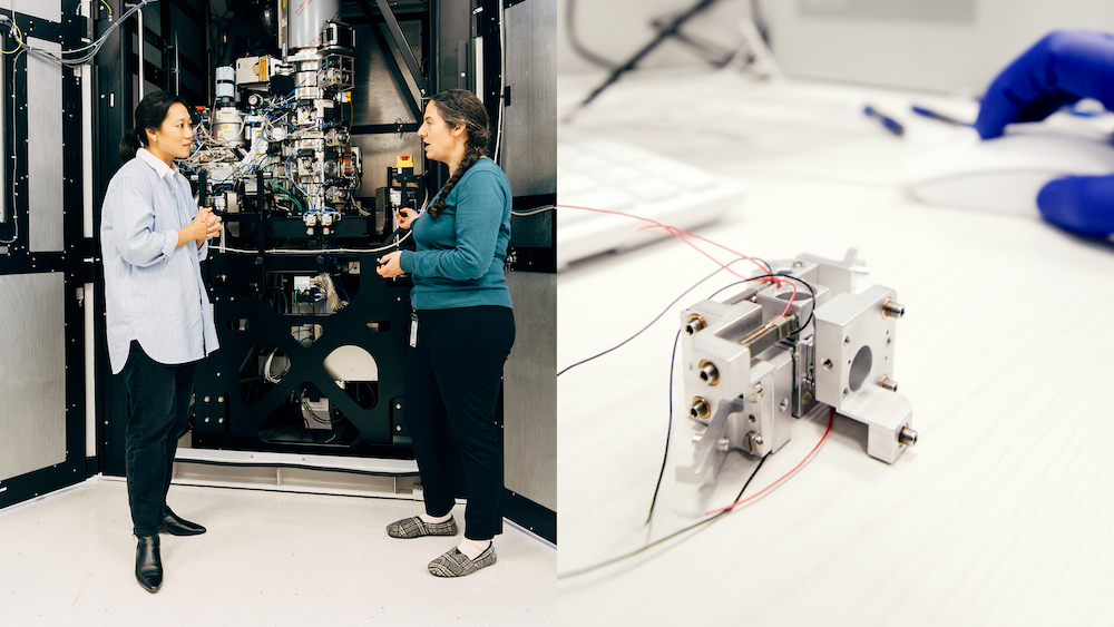
From the outside, both configurations of the laser phase plate–enhanced microscope look much like other cryo-electron microscopes. That’s because the entire laser apparatus fits inside a device the size of an espresso cup, which sits inside the microscope column.
But Biohub’s dual-laser design, installed in a custom Thermo Scientific Krios microscope, was engineered to work reliably enough that others can eventually reproduce it — a necessary condition before any technology moves from a single lab into the broader research community. The parallel development compressed what might have taken decades into seven years, with each team specializing while sharing results across the collaboration.
In three papers — one published in Science, demonstrating enhanced contrast for small proteins with the single-laser phase plate; one published in Nature Communications laying out the theoretical framework for the xLPP; and one a preprint describing Biohub’s design and implementation of the xLPP — researchers at UC Berkeley and Biohub report that the laser phase plate, in both the single- and dual-laser configurations, is not just theoretically elegant. It works in a modern cryo-EM, and it significantly improves contrast in images of small proteins.
In a series of paired experiments, the Berkeley team first imaged aldolase, a medium-sized enzyme that serves as a standard benchmark in cryo-EM, and then hemoglobin, the oxygen-carrying protein in red blood cells. With a molecular weight of just 64 kilodaltons, hemoglobin sits near the lower frontier of what conventional cryo-EM can resolve, right at the edge of what the field can barely see.
When the first real results came in, Müller’s team didn’t celebrate but went quiet. Technician Jonathan Remis spent a weekend running the images through the software pipeline. “He just goes silent for two days,” recalls Jessie Zhang, a postdoc in the Müller lab and lead author of the Science paper. “We’re just, like, how’s the data going? Is it bad? Then he’s like, ‘What if it’s so good I want to make sure it’s correct before sharing it with you?’”
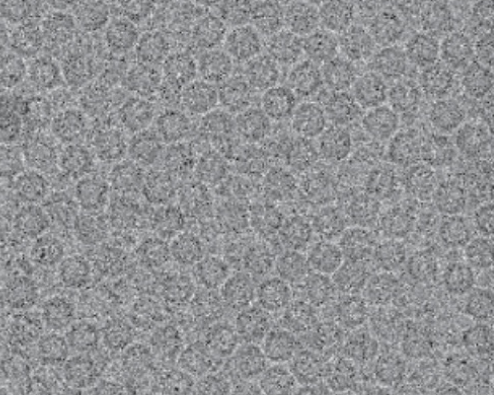
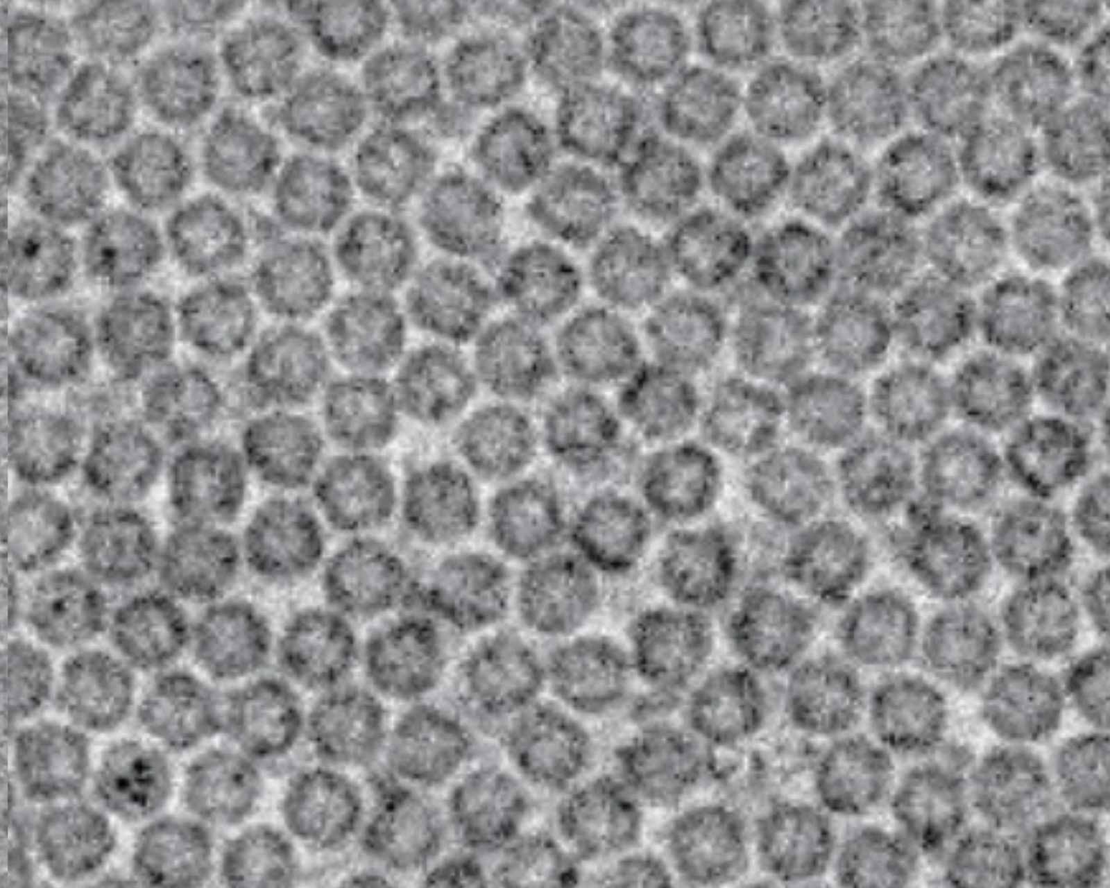
The meticulous analysis was worth it. With the laser off, the images looked like every other cryo-EM image: usable, but constrained. With the laser on, resolution improved by up to 44%. The proteins sharpened. Features that had been blurry became clearly visible.
Meanwhile, working at Biohub, Carragher and her team used the dual laser phase plate to image apoferritin — a small iron-storage protein — at 1.8 angstroms, approaching the theoretical resolution limit of the technology. They also imaged frozen E. coli bacteria, demonstrating that the laser significantly boosted contrast in conditions where conventional imaging falls short.
These early, proof-of-principle results on purified proteins and bacteria establish that the phase plate works, reproducibly, in a real microscope. Now, researchers at Biohub and UC Berkeley are testing the Biohub technology on the complex, intact cells where the technology is ultimately expected to make its biggest mark, through cryo-ET.
Over the last decade, cryo-ET has revealed glimpses of biology no other method can access: ribosomes bound to antibiotics inside bacteria, nuclear pore complexes in their native context, and the machinery that drives cell division. But cryo-ET has always been constrained by the same contrast problem; more so, in fact, because tomographic samples are thicker, messier, and more complex.
A phase plate that reliably boosts contrast inside cells would be transformational for the field. “The real promise is tomography,” says David Agard, Biohub’s founding scientific director of imaging.
“When you can see inside a cell with phase contrast — when you can actually watch proteins interact with other proteins in context — that’s when biology starts to come alive.”
— David Agard, founding scientific director of imaging at Biohub
Both teams are pursuing the laser phase plate’s benefits for cryo-ET. “Cryo-ET right now is in demonstration mode, not experimentation mode, because everything is so slow,” Carragher says. True use of the technique, she explains, will mean a researcher can bring a biological question to the microscope, spend some weeks collecting and analyzing data, and iterate toward an answer — not spend years staggering toward a single structural characterization.
The laser phase plate is the field’s best current candidate for that upgrade, Carragher says. Better contrast also allows researchers to correct for much of the noise inherent in tomographic data, the same way improved cameras unlocked single-particle cryo-EM a decade ago. “Thanks to the tremendous work of Biohub, routine tomography with the laser phase plate is now within close reach,” says Müller.
Biohub is also working to ensure the technology’s benefits extend to the global scientific community. All its tomography data — including tens of thousands of annotated tomograms — are freely shared with the research community through the CryoET Data Portal, with the aim of accelerating the entire cryo-ET pipeline for labs worldwide.
Biohub has committed significant resources to the laser phase plate over the past seven years not because success was guaranteed, but because the potential upside was large enough to justify the risk. “Too often, promising technologies get stuck between proof-of-concept and real-world deployment — the ‘valley of death,’” says Otte. “We decided that this was too important to let that happen.”
The strategy was never just about funding a single lab. It was a complex, years-long collaboration with partners across engineering, optics, software, and electron microscopy. The laser phase plate is core to Biohub’s larger scientific ambition of building the technologies needed to accelerate science that cures or prevents disease.
In April, Biohub announced the Virtual Biology Initiative: a five-year, $500-million commitment to generating new cellular datasets to train AI models, spanning from the molecule to the organism, in both health and disease. The laser phase plate will be critical to obtaining the detailed images needed to train these models.
The laser phase plate, in this context, is a significant step toward making the cell’s interior readable by researchers and AI alike. The destination — atomic-resolution imaging inside intact cells, the full molecular atlas of health and disease — remains ahead. But a new tool to get there is, for the first time, real.
此内容由惯性聚合(RSS阅读器)自动聚合整理,仅供阅读参考。 原文来自 — 版权归原作者所有。